![[Translate to English:] Organisme partenaire](/fileadmin/_processed_/0/1/csm_CNRS_3a745ef80f.png "[Translate to English:] Accès à l'un des organismes partenaires : CNRS")
![[Translate to English:] Organisme partenaire](/fileadmin/_processed_/f/f/csm_1200px-Inserm.svg_e391664898.png "[Translate to English:] Accès à l'un des organismes partenaires : INSERM")
![[Translate to English:] Organisme partenaire](/fileadmin/_processed_/b/b/csm_chu_25c6c9f960.png "[Translate to English:] Accès à l'un des organismes partenaires : CHU Lille")
![[Translate to English:] Organisme partenaire](/fileadmin/_processed_/6/1/csm_pasteur_21b9760e1f.png "[Translate to English:] Accès à l'un des organismes partenaires : Institut Pasteur de Lille")
Principle
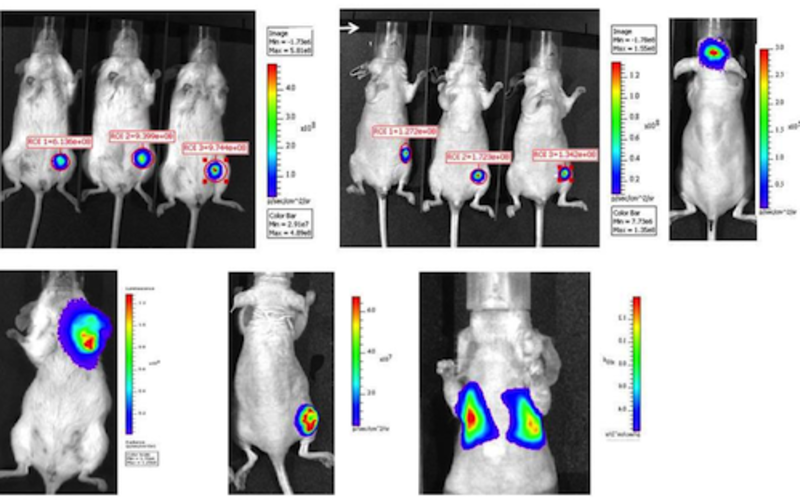
In vivo imaging is the non-invasive visualization of living organisms for research or diagnostic purposes. There are generally two key areas: anatomical/morphological imaging and molecular imaging. In molecular imaging, cellular functions or molecular processes are visualized, usually using biomarkers. In anatomical imaging, no markers are used and visualization is based on the intrinsic properties of the tissues and organs to be observed, such as X-ray attenuation in computed tomography. Molecular imaging very often uses biomarkers labeled by bioluminescence or fluorescence.
With in vivo molecular imaging, several molecular events can be monitored simultaneously, for example visualizing drug effects, to optimize drug and gene therapy, to monitor tumor growth or to study disease progression in living animals and plants. Insights into transcriptional and translational feedback loops, which are influenced by environmental signals such as light or temperature, even allow observation of biological clocks in plants. Compared to other methods of measuring molecular processes, in vivo imaging has several major advantages:
- It provides the spatial location of the process or molecule.
- It is dynamic: changes over time can be tracked.
- It can be repeated several times in the same individual.
- It allows to observe a (mostly) undisturbed process, i.e. the results are physiologically relevant.
These advantages make in vivo imaging a very useful technology in many research and diagnostic areas.