![[Translate to English:] Organisme partenaire](/fileadmin/_processed_/0/1/csm_CNRS_3a745ef80f.png "[Translate to English:] Accès à l'un des organismes partenaires : CNRS")
![[Translate to English:] Organisme partenaire](/fileadmin/_processed_/f/f/csm_1200px-Inserm.svg_e391664898.png "[Translate to English:] Accès à l'un des organismes partenaires : INSERM")
![[Translate to English:] Organisme partenaire](/fileadmin/_processed_/b/b/csm_chu_25c6c9f960.png "[Translate to English:] Accès à l'un des organismes partenaires : CHU Lille")
![[Translate to English:] Organisme partenaire](/fileadmin/_processed_/6/1/csm_pasteur_21b9760e1f.png "[Translate to English:] Accès à l'un des organismes partenaires : Institut Pasteur de Lille")
Principle
One of the challenges of optical imaging in biology is to visualize biological structures and organisms in three dimensions under conditions that preserve their three-dimensional integrity. However, the phenomena of scattering and absorption of light, due to the thickness of the sample, appear and are an intrinsic obstacle to the in-depth exploration of these samples. To meet the needs of 3D imaging of biological samples, a large number of techniques of sample transparisation have been developed, some based on organic solvents, others based on aqueous solutions, with different properties and various applications
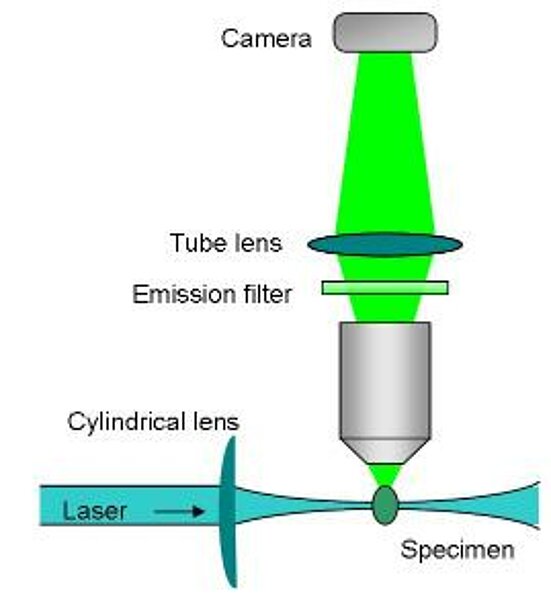
Light sheet microscopy is based on a simple principle. The light that illuminates the observed biological object is no longer a cylindrical laser beam, but a sheet of light a few micrometers thick. In an original way, this sheet of light is positioned perpendicular to the objective which will detect the fluorescence emitted by the object. Thus, as on a virtual slicer, the displacement of the object in this sheet of light will allow to carry out optical cuts at all the depths of the sample, thus allowing to observe it in its totality.

Instrument available :

Campus HU
Description :
- Marque : LaVision Biotec
- Type de statif : Droit
- Logiciel : Imspector
- Lasers (nm) : 488 / 561 / 633 / 785
- Modalité : Microscope à feuille de lumière
- Détecteur : Caméra sCMOS Andor Neo
- Objectifs : 2x/0.5 (Multi)| 4/0.3 (Multi) | 12x/0,53 (Multi) | 20x/0,95 (DBE) | 2x Zoom
- Platine : Motorisée
- Supports : Porte-échantillon
- Z-Stacks : Oui
- Mosaïques : Oui
- Multi-positions : Oui
- Accessoires : Illumination Gauche+Droite
Applications :
- Observation 3D d'organes transparisés DISCO, CUBIC,TDE,...
- Détection simultanée de 4 couleurs de 500 nm à 850 nm.
- Acquisitions automatisées multi-position et mosaïque.
Localisation : Faculté de Médecine - Pôle Recherche 4ème étage
Contact : Meryem Tardivel | Antonino Bongiovanni | 03.20.62.34.93