![[Translate to English:] Organisme partenaire](/fileadmin/_processed_/0/1/csm_CNRS_3a745ef80f.png "[Translate to English:] Accès à l'un des organismes partenaires : CNRS")
![[Translate to English:] Organisme partenaire](/fileadmin/_processed_/f/f/csm_1200px-Inserm.svg_e391664898.png "[Translate to English:] Accès à l'un des organismes partenaires : INSERM")
![[Translate to English:] Organisme partenaire](/fileadmin/_processed_/b/b/csm_chu_25c6c9f960.png "[Translate to English:] Accès à l'un des organismes partenaires : CHU Lille")
![[Translate to English:] Organisme partenaire](/fileadmin/_processed_/6/1/csm_pasteur_21b9760e1f.png "[Translate to English:] Accès à l'un des organismes partenaires : Institut Pasteur de Lille")
Principle
In the field of research, the use of automated systems makes it possible to facilitate, accelerate and standardize the acquisition and quantification of immunofluorescence, immunohistochemistry or contrast.
Within the BICeL, you have the possibility to automate your plate and slide acquisitions.
The scanners are made up of components similar to those of the most powerful motorized microscopes, but the eyepieces and the manual control tools for positioning and focusing have been removed. It has a single aperture that can accommodate up to a hundred samples. It generally uses a 20x objective with a large numerical aperture allowing a very good resolution of the final images. It is also possible to install a real 40x lens. They are therefore particularly adapted to users with large volumes of slides and dedicated and standardized digitization problems. These systems help scientists to evaluate the molecular expression of markers, their location within their sample and to standardize the score attributed to the signal intensity observed on slides.
The impossibility of long-term preservation of immunofluorescent markings disappears thanks to this technology which allows not only the digitization of an entire immunofluorescence image in a few minutes, but also its almost indefinite preservation.
Available instruments:
Campus HU
Description :
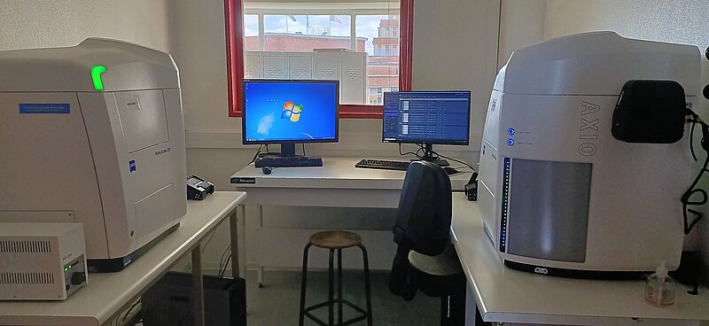
- Marque : Zeiss Axioscan Z1
- Type de statif : Droit
- Logiciel : Zen Blue
- Source Colibri Fluo (nm) : 405 / 488 / 561 / 633
- TL : BF
- Modalité : Microscope champs-large
- Détecteur : Caméra Orca Flash 4 (Fluo) | Caméra Hitachi HV F202 (Visible)
- Objectifs : 10x/0.3 | 20/0.8 | 40x/0,9
- Platine : Motorisée
- Supports : Chargeur 100 lames
- Z-Stacks : Oui
- Mosaïques : Oui
- Multi-positions : Oui
- Accessoire : 25 supports de lames 26 x 76 mm | 10 supports de lames 52 x 76 mm
Applications :
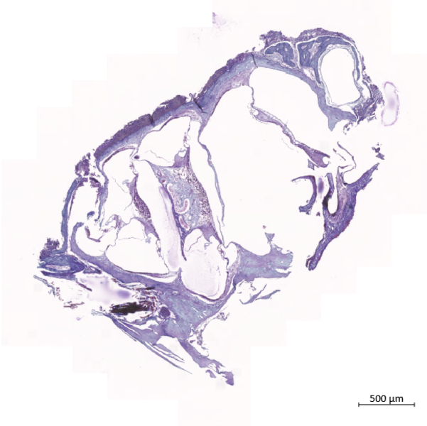
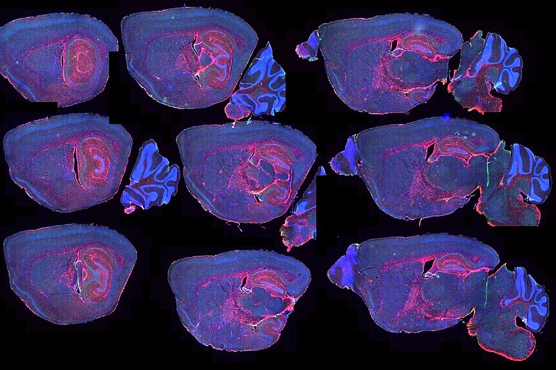
- Observation 2D plein champs de coupes histologiques IF ou IHC.
- Observation de l'échantillon par le dessus.
- Détection simultanée de 4 couleurs de 400nm à 750nm.
- Acquisitions automatisées
- Corrections automatisées (Ombrage et balance des blancs)
Localisation : Faculté de Médecine - Pôle Recherche 4ème étage
Contact : Meryem Tardivel | Antonino Bongiovanni | 03.20.62.34.93
Tarif : 12€/heure
Description :
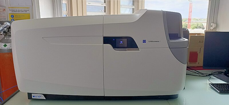
- Marque : Zeiss Cell Discoverer 7
- Type de statif : Inversé
- Logiciel : Zen Blue
- Source Colibri Fluo (nm) : 405 / 458 / 488 / 514 / 561 / 633
- TL : BF / Ph / Gradient
- Modalité : Microscope champs-large
- Détecteur : Caméra Orca Flash 4 (Fluo) | Caméra Zeiss 506 (Fluo)
- Objectifs : 5x/0.35 | 20/0.95 | 40x/0,9 | 50x/1.2 (Water)
- Lentille : 0.5x | 1x | 2x
- Platine : Motorisée
- Chambre d'incubation Temp. et CO2 : Pecon
- Supports : Plaques / Plaques multipuits / Lames
- Z-Stacks : Oui
- Mosaïques : Oui
- Multi-positions : Oui
- Accessoire : Désinfection UV
Applications :
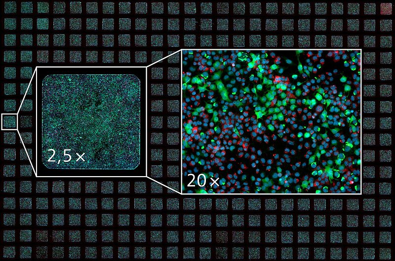
- Observation plein champs de culture 2D ou 3D
- Observation de l'échantillon par le dessous.
- Détection simultanée de 6 couleurs de 400nm à 750nm.
- Acquisitions automatisées
- Détection automatique du format de plaque
- Injection en cours d'expérience de liquide
Localisation : Faculté de Médecine - Pôle Recherche 4ème étage
Contact : Meryem Tardivel | Antonino Bongiovanni | 03.20.62.34.93