![[Translate to English:] Organisme partenaire](/fileadmin/_processed_/0/1/csm_CNRS_3a745ef80f.png "[Translate to English:] Accès à l'un des organismes partenaires : CNRS")
![[Translate to English:] Organisme partenaire](/fileadmin/_processed_/f/f/csm_1200px-Inserm.svg_e391664898.png "[Translate to English:] Accès à l'un des organismes partenaires : INSERM")
![[Translate to English:] Organisme partenaire](/fileadmin/_processed_/b/b/csm_chu_25c6c9f960.png "[Translate to English:] Accès à l'un des organismes partenaires : CHU Lille")
![[Translate to English:] Organisme partenaire](/fileadmin/_processed_/6/1/csm_pasteur_21b9760e1f.png "[Translate to English:] Accès à l'un des organismes partenaires : Institut Pasteur de Lille")
Transmission electron microscope
Principle
Transmission electron microscopy (TEM) involves imaging an ultrathin sample (natural or "slice" obtained by ultramicrotomy) by means of a dilated electron beam that passes through the entire sample at the same time.
Instruments available:
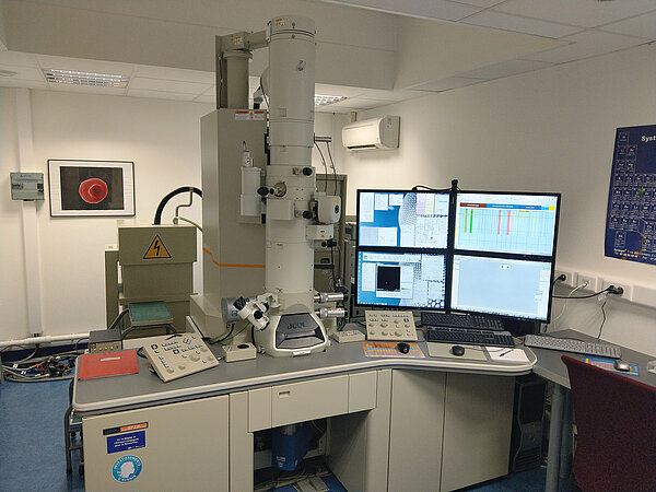
Campus CS
Description
- Brand : JEOL
- Model : JEM-2100
- Type : Microscope électronique en Transmission
- Electron source : LaB6 filament
- High voltage : 200kV
- Pole piece : Cryo
- Maximum resolution: image mode 0.14nm, point mode 0.27nm
- Max magnification: x 1.000.000
- 2 cameras (Gatan): SC200D, US4000
- STEM detector (JEOL) : Bright field, Dark Field
- EDX detector (Bruker) : XFlash 5030
- EELS, EFTEM energy filter (Gatan): GIF Quantum
- MDS "minimum dose system" (JEOL)
- Anti-contamination system (ACD)
- Motorised sample displacement with magnification control
- Magnification: up to x 1000.000
- Sample holders (JEOL): Standard, Beryllium (EDX), Quartet, Electron Tomography
- Sample holders Cryo + temperature controller (Gatan)
- Software: JEOL (TEM /STEM settings, STEM acquisition), Gatan DigitalMicrograph GMS 2 (TEM image acquisition, tomography, EELS, EFTEM...), Bruker Esprit 2 (EDX analysis)
Applications
- TEM (Transmission Electron Microscopy) observations: Bright field, Dark Field
- STEM (Scanning Transmission Electron Microscopy) observations: Bright field, Dark Field, HAADF
- Electron Tomography (Gatan software)
- Cryo-observation
- Chemical analysis: EDX / EELS
Location: Cité Scientifique - Building SN3 - Basement
Contact : Loïc Brunet | 03.20.43.41.03
Rate of use
- 54€/hour " for internal academics " (supply of liquid nitrogen in addition)
- 67€/hour "for external academics" (liquid nitrogen supply not included)
- Sample preparation (on quotation)
- Rates for all services for private companies (on request)